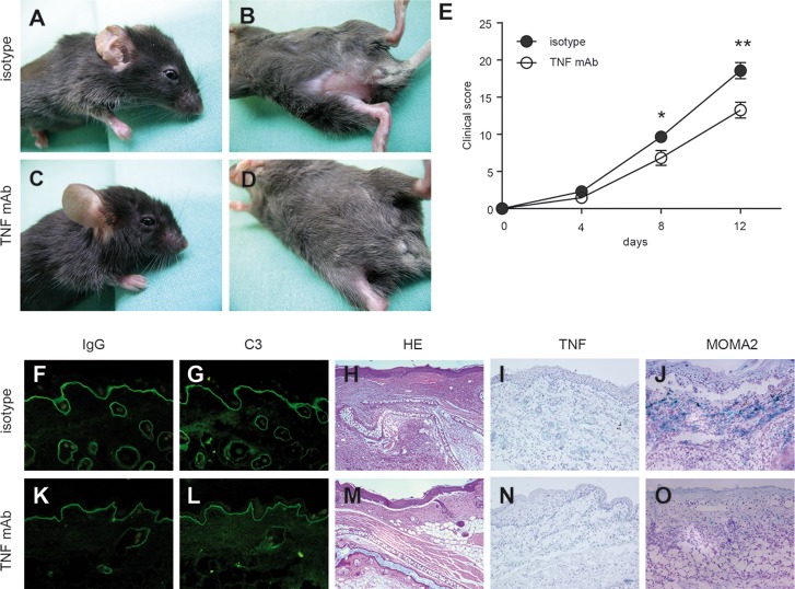
Figure 2.
Prophylactic TNF blockade reduced the extent of skin disease in antibody transfer–induced EBA. Mice injected with rabbit anti-mCOL7C IgG and treated with (A,B) an isotype antibody (n = 8) or (C,D) a monoclonal antibody to TNF (TNF mAb, n = 6) 2 d prior to initial anti-mCOL7C IgG injection, followed by five treatments every other day until d 8. Ears and trunks of isotype-treated mice demonstrated erythema and erosions, while less disease was observed in TNF mAb–treated mice. (E) Time course of clinical disease development, shown as the percentage of body surface area affected by blistering (*p < 0.05, **p < 0.01). Representative ear samples from (F–J) isotype-treated and (K–O) TNF mAb–treated mice with EBA were analyzed histopathologically and immunologically. By direct immunofluorescence microscopy, similar (F,K) IgG and (G,L) C3 deposits were observed at the dermal-epidermal junction in both experimental groups. Histopathological analysis of lesional skin demonstrated comparable (H,M) dermal-epidermal separation in both treatment groups, but (I,N) TNF and (J,O) MOMA-2 were detected to a lesser extent in (N,O) TNF mAb–treated mice compared with (I,J) isotype-treated mice. Magnification of all sections is 200×.

