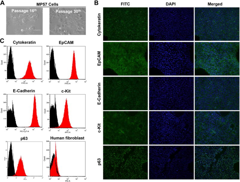
Figure 1.

Morphology and characterization of MP57 primary cells. (A) Photographs of MP57 morphology at passage 16th and 30th, taken under a light inverted microscope (magnification 40×). (B) Immunofluorescence staining of thymic epithelial markers in MP57. The staining of cytokeratin clone AE1/AE3, EpCAM, E-Cadherin, c-Kit, and p63 are indicated by FITC. DAPI was used for staining of nuclei. (C) Cell surface marker analysis of MP57 by flow cytometry. Cytokeratin clone AE1/AE3, EpCAM, p63, c-Kit, and E-Cadherin, and human fibroblast antibody were analyzed. Black histogram represents control isotypes.
