Figure 2.

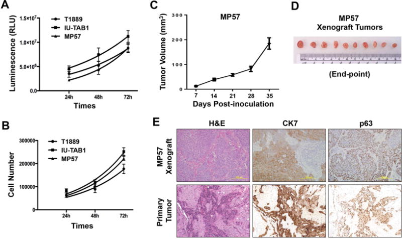
Proliferative and tumorigenic activities of MP57 cells. Cell proliferation was measured by (A) CellTiter Glo Viability assay, and (B) Trypan-Blue dye exclusion method. For comparison, T1889 and IU-TAB1 cells were also examined. (C) Tumorigenicity of MP57 cells in immunocompromised athymic nude mice (n=10). The curve represents the growth of subcutaneous tumors within the 5-week window. (D) MP57 xenograft tumors at the experimental end-point (day-35 post-inoculation). (E) Histological comparison between MP57 xenograft tumor and the primary tumor obtained from patient autopsy. Morphology of tumor cells was examined by H&E staining. The expression of cytokeratin 7 and p63 was determined by immunohistochemistry.
