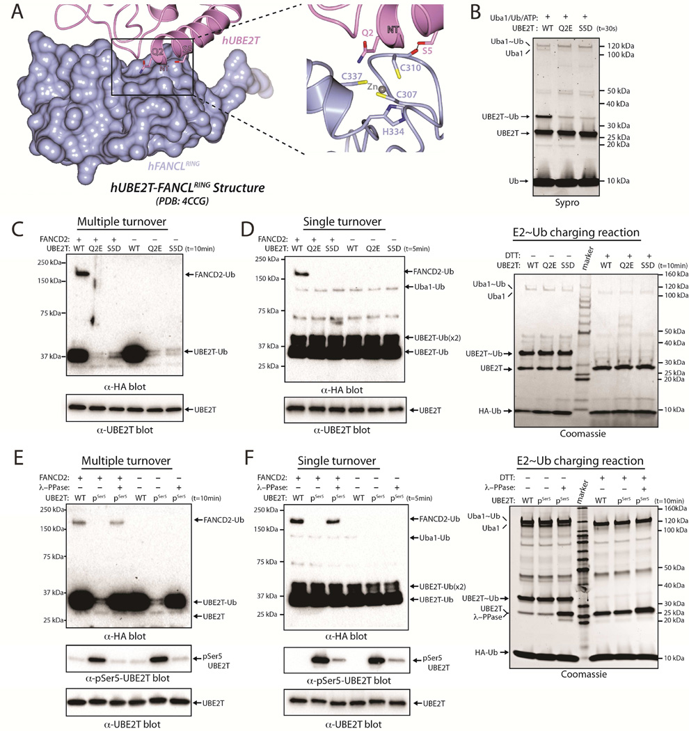
Figure 6. Acidity at the N-terminus of E2 negatively regulates FANCL-mediated UBE2T ubiquitination of FANCD2.
(A) Structure of hUBE2T (slate) in complex with the FANCL RING domain (rose) (PDB: 4CCG), with selected residues shown as sticks.
(B) E1-E2 thioester transfer assays of the indicated proteins.
(C) FANCL-mediated UBE2T monoubiquitination of FANCD2 performed under multiple turnover conditions (top). UBE2T loading control (bottom).
(D) FANCL-mediated UBE2T monoubiquitination of FANCD2 performed under single turnover conditions (left). E2~Ub charging control (right).
(E) Multiple turnover FANCD2 monoubiquitination assay as in C (top). E2s were treated or untreated with lambda phosphatase, as indicated. Anti-pSer5 UBE2T western blot (bottom).
(F) Single turnover FANCD2 monoubiquitination assay as in D (top). Anti-pSer5 UBE2T western blot (bottom).

