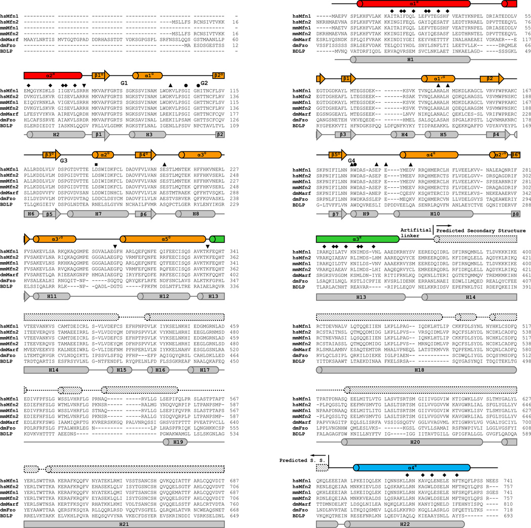
Extended Data Figure 3. Sequence alignment of mitofusins and BDLP.
Sequence alignment of mitofusins and BDLP. Amino acid sequences of human (hs) Mfn1 (UniProt accession Q8IWA4) and Mfn2 (O95140), mouse (mm) Mfn1 (Q811U4) and Mfn2 (Q80U63), fruit fly (Drosophila melanogaster, dm) Marf (Q7YU24), fruit fly Fzo (O18412) and the bacterial dynamin-like protein (BDLP) from Nostoc punctiforme (B2IZD3) are aligned using Clustal W44. Residues with a conservation of 100% are in red shades, greater than 80% in green shades and 50% in grey shades, respectively. α-helices are shown as cylinders and β-strands as arrows for both nucleotide-free human Mfn1 (above the sequences) and nucleotide-free BDLP (2J,under the sequences). In the case of human Mfn1, the secondary structure signs are coloured as in Fig. 1b and labelled as in Figs. 1b–d for Mfn1IM regions. Secondary structural elements of the missing HD2 and TM predicted from the PHYRE2 server45 (exclusively α-helices) are depicted as shaded cylinders with dashed outlines. For BDLP, the secondary structure signs are coloured grey and labelled according to the previous report21. The G1-G4 elements are specified in the sequences. Key residues on human Mfn1 are also indicated, including those involved in the hydrophobic core of HD1 (♦), Hinges (▼), guanine nucleotide binding and hydrolysis (●), G interface (▲), and the plausible HD1-HD2 conformational change (■).

