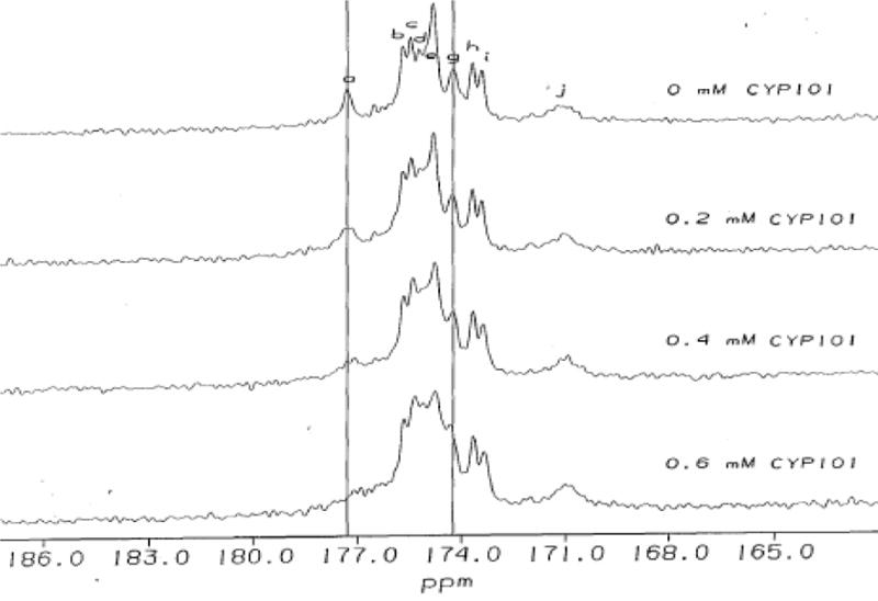
Figure 3.

Changes in 13C NMR spectra of 13C labeled Pdx° upon addition of 0.2 mM increments of CYP101°. Residues in paramagnetic region of Pdx° affected by complexation are labeled. Peak labels a,b,h,i,j in 13C spectrum correspond to cysteine residues 39, 85, 86, 48 and 45 respectively; d,g correspond to serine residues 44 and 42 respectively; c,e,f correspond to glycine residues 41, 37 and 40 respectively. Differential changes in line-broadening and chemical shift for Cys39 and Ser42 are indicated by vertical lines.
