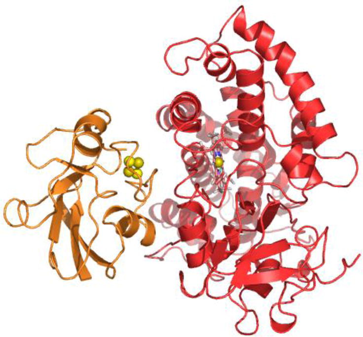
Figure 7.

Ribbon representation of the NMR structure of Pdx°/CYP101° complex derived from docking of Pdx° (in orange) and CYP101° (in red) structures in HADDOCK56 using RDC orientational and intermolecular paramagnetic spin label distance restraints. The [2Fe-2S] metal cluster of Pdx° is shown as spheres, while the heme prosthetic group of CYP101° is shown as sticks. The figure was prepared using the program PyMOL.62
