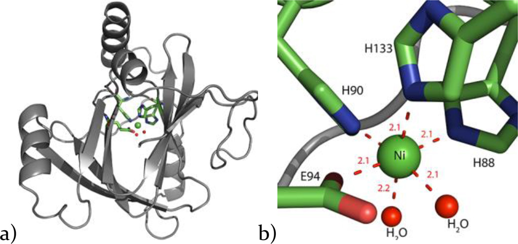
Figure 3. X-ray crystal structure of Ni-MmARD.
a) The X-ray crystal structure of Ni-MmARD with the Ni atom shown as a green sphere, b) The active site with the Ni atom as a green sphere and its protein ligands H88, H90, H133, E94 represented as sticks and two water molecules shown as red spheres forming an octahedral coordination geometry. The metal co-ordination distances are shown as red dotted lines with distances measured in Å.

