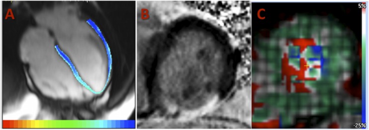
Figure 2.
Images from the CMR protocol in the DCCT/EDIC study. A: T1 mapping assessment of the LV from a four-chamber view using a TI scout Look-Locker sequence. B: LGE image in a short-axis view of the LV (presence of transmural scar in the inferior wall). C: Assessment of LV deformation (strain map) from tagging sequences in a midventricular short-axis view.

