
Figure 2.
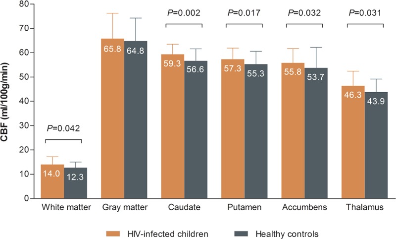
HIV-infected children have higher CBF in white matter, basal ganglia, and thalamus. We compared ASL-measured CBF between HIV-infected children and healthy controls using linear regression analysis adjusted for age (>16 years), sex, and hematocrit. Prior to analysis, CBF values for caudate nucleus, putamen, nucleus accumbens, and thalamus were normalized subject-wise using the overall mean gray matter CBF. The error bars represent standard deviations. ASL = arterial spin labeling, CBF = cerebral blood flow, HIV = human immunodeficiency virus.