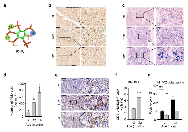
Fig. 1.
Increased numbers and dysregulation of bone marrow (BM)-derived macrophages (BMDMs) in aged mice. (A) Schematic illustration of IC-IP6. (B) IC-InsP6 accumulation in the liver was visualized by double-staining with Prussian blue (iron) and the macrophage-specific anti-F4/80 antibody (brown). Scale bars: top, 50 μm; bottom, 20 μm. (C) Prussian blue staining for assessment of IC-IP6 uptake by BMDMs in decalcified femur sections from age-matched mice. (D) Quantification of Prussian blue-positive cells per mm2 in the BM of mice intravenously injected with 10 μM IC-IP6, mean ± SE (n = 10, ***P < 10−3). (E) Immunohistochemical staining of F4/80 in decalcified femur sections from age-matched mice. Scale bars: top, 200 μm; bottom, 50 μm. (F) Frequency of total BMDMs assessed by flow cytometric analysis of CD11b+MHCII+F4/80+ cells in the BM of age-matched mice, mean ± SE (n = 6–7, ***P < 10−3). (G) M1 and M2 macrophage polarization assessed by flow cytometric analysis of CD11b+/CD206+ and CD11b+/CD206− macrophages from MHCII+F4/80+ cells in BM cells of age-matched mice, respectively, mean ± SE (n = 6–7, **P < 0.01).

