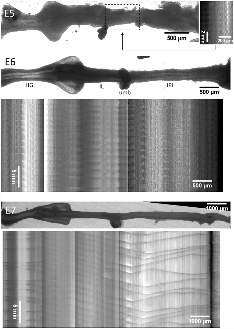
Fig 2. Representative motility patterns at E5, E6 and E7 (S2–S4 Videos).
At E5, the motiligram is deduced from the sole beating region of the gut which is the distal jejunum (dashed box). At later stages, the motiligram is placed right below the region of the gut from which it was derived; spatial scales are the same for still images of the gut and motiligrams. The main anatomical segments of the gut are indicated at E6, HG: hindgut, IL: ileum, JEJ: jejunum, umb: umbilicus.

