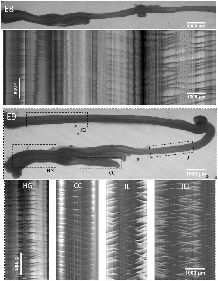
Fig 3. Representative motility patterns at E8 and E9 (S5 and S6 Videos).
The E8 motiligram is placed right below the region of the gut from which it was derived; at E9, separate motiligrams were derived from regions indicated with dashed boxes. HG: hindgut, IL: ileum, JEJ: jejunum, umb: umbilicus, CC: caecal appendix.

