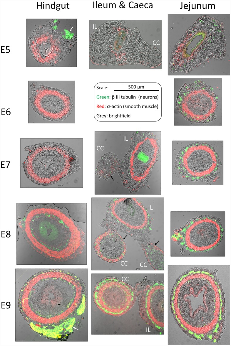
Fig 5. Immunohistochemical staining of neurons (green, β-III tubulin) and smooth muscle (red, α-actin) in the hindgut, jejunum (pre-umbilical midgut) and ileum (IL)—caeca (CC) region from E5 to E9, transverse sections.
The brightfield image is overlaid in grey. The scale bar is the same for all sections. The enteric nervous system appears as rings of neurons located around the circular smooth muscle layer; concentrated, non-axisymmetric patches of neurons visible on the periphery of sections E5-HG and E9-HG are the extrinsic innervation (white arrows). The epithelium is visible on the brightfield image, but sometimes stains non-specifically either to Alexa488 (e.g., E7-IL, E8-HG) or to CY3 (e.g., E8-JEJ) secondary antibody.

