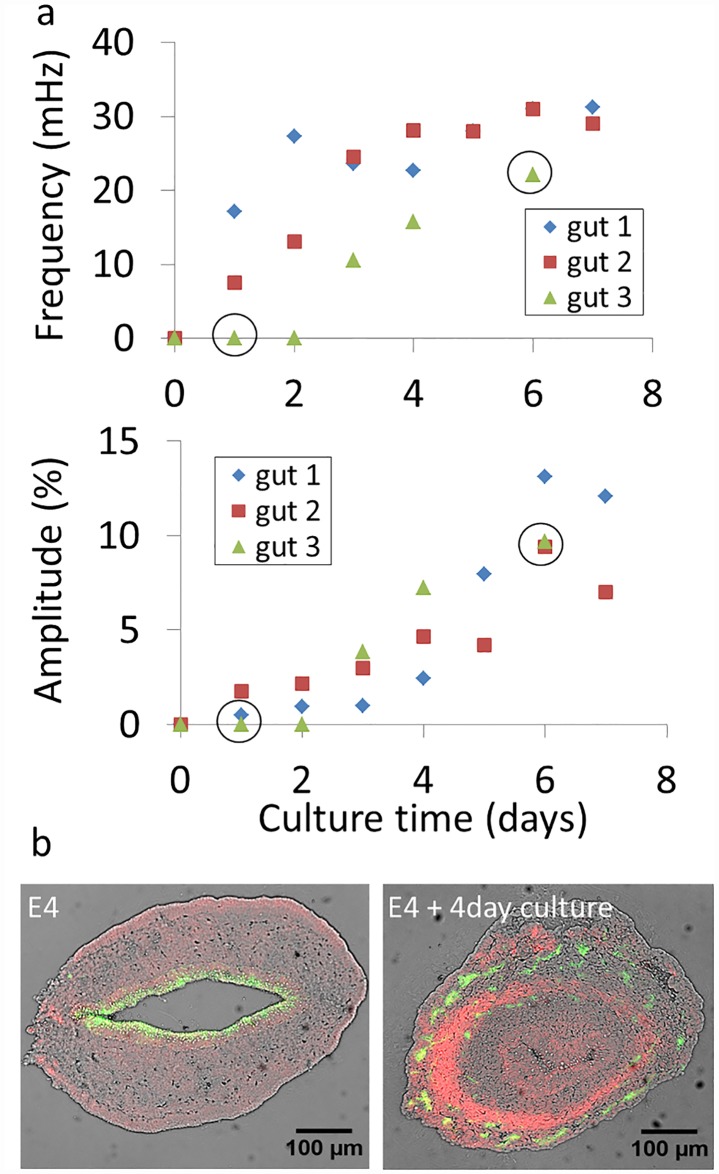
Fig 7.
a) Evolution of jejunal contractile wave frequency and amplitude of E4 guts (n = 3) kept up to 7 days in culture. Circled data points indicate that the full time-lapse video corresponding to this condition is available as S9 & S10 Videos. b) Immunohistochemical staining of neurons (green, β-III tubulin) and smooth muscle (red, α-actin) in native E4 midgut (left) and E4 midgut kept 4 days in culture (right). The brightfield image is overlaid in grey. For the native E4 midgut, the epithelium stains weakly and non-specifically to Alexa488.

