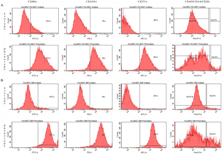
Fig 3. MSC characterization by flow cytometry.
A) Wharton’s jelly mesenchymal stem cell (WJMSCs) and, B) bone marrow mesenchymal stem cell (BMMSCs). All MSCs stained positive for CD90 by fluoroscein isocyanate (FITC), CD105 by phycoerythrin (PE) and CD73 by allophycocyanin (APC); and they were negative for hematopoietic markers CD45, CD34, CD14 or CD11b, and CD20 as analyzed by Cell Profiler (CP) software (Broad Institute).

