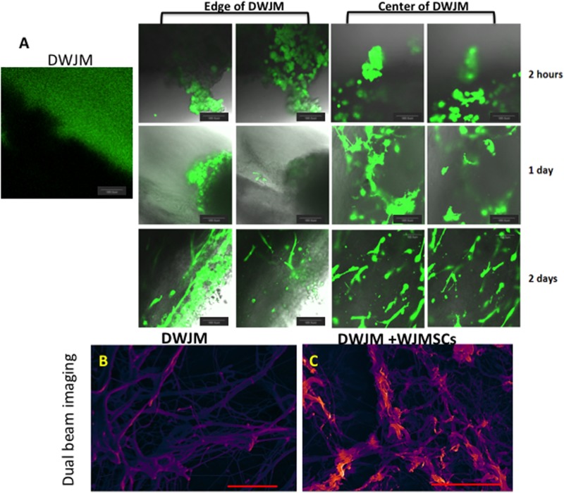
Fig 4. Transplantation and culturing of WJMSCs on DWJM.
A) Confocal microscopy images of DWJM and WJMSCs on DWJM after 2 hours (upper panel), 1 day (center panel), and 2 days (lower panel) post- cell seeding. The cells are labeled with calcein acetylmethyl (AM) that stains the live cells in green. Dual beam imaging of B) DWJM and C) DWJM seeded with WJMSCs for 1 week. The Everhart-Thornley detector (ETD) is a standard secondary electron detector used in scanning electron microscopy to study topography, while the circular backscatter (CBS) is a backscatter detector that reveals lipid content when samples are stained with osmium tetroxide (OT) (red/orange). Images have been pseudo-colored to enhance definition proportional to secondary electron signal for ETD. (Scale bar is 20 μm.) DWJM appears to be a fibrous interpenetrating network with varying pore sizes, while WJMSCs were arranged along the fibers of DWJM.

