Figure 2.
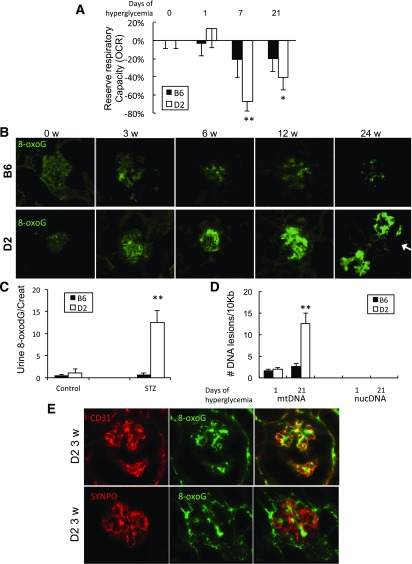
Decreased mitochondrial function and increased mtDNA damage in glomeruli of diabetic D2 mice. A: Mitochondrial respiratory reserve capacity was measured by OCR (OCR uncoupled respiration [FCCP] over basal respiration) of isolated glomeruli from B6 and D2 controls, STZ-B6, and STZ-D2 mice with diabetes for the days indicated (values are mean percentage reduction in OCR ± SEM; n > 6 mice/time point). B: Immunofluorescent detection of oxidative DNA marker 8-oxoG in glomeruli from B6 (top) and D2 mice (bottom) with 3, 6, 12, and 24 weeks of STZ-induced diabetes and untreated control mice 0 weeks. Arrow indicates 8-oxoG–positive staining in tubular cells. C: Urine 8-oxodG (nmol) relative to urine creatinine (Creat) in B6 and D2 control mice and 3-week diabetic STZ-B6 or STZ-D2 mice. D: Quantification of lesion frequencies in mtDNA and nuclear DNA (nucDNA) by quantitative PCR in isolated glomeruli of B6 and D2 mice after 1 and 21 days of hyperglycemia (n = 6; ±SEM) relative amplification normalized to nondamaged (0 days controls). E: Representative images of double immunofluorescence staining detecting endothelial cell marker CD31 (red, top left), 8-oxoG (green, top middle), and merge (top right) or podocyte marker synaptopodin (SYNPO; red, bottom left), 8-oxoG (green, bottom middle), and merge (bottom right) in glomeruli of 3-week diabetic STZ-D2 mouse. *P < 0.05, **P < 0.01 versus untreated control mice.

