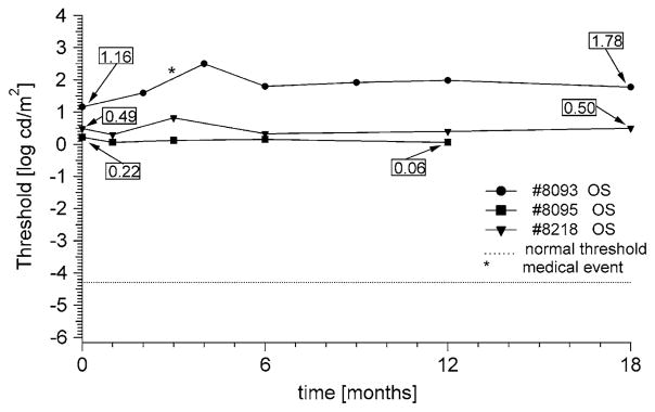
Fig. 5.
Longitudinal D-FST data from a male (#8093) and two females (#8095, #8218) with isolate RP. Their visual acuity was limited to LP only, and both rod and cone full-field ERGs were non-detectable (<2.0 and <0.1 μV, respectively). The star denotes an episode of viral conjunctivitis. There was an increase in threshold of 0.6 log unit over 18 months for #8093, while visual acuity measurements remained at LP. Patients #8095 and #8218 remained stable within the 0.3 log confidence range

