. 2017 Feb 21;6:e20420. doi: 10.7554/eLife.20420

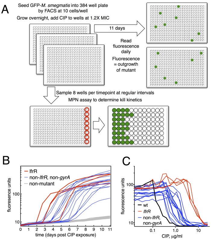
Figure 1. Selection of mutants at low ciprofloxacin concentration in 384 well plates.

(A) Schematic of the selection strategy. Starting at t = −16 hr, 10 M. smegmatis cells are introduced into wells of three 384 well microtiter plates containing 15 µl of growth medium and grown overnight at 37°C. At t = 0, 15 µl of medium containing CIP are added to yield a final CIP concentration of 0.3 µg/ml. Plates were sealed with foil and placed at 37°C. At t = 0 and at roughly daily intervals afterwards, plates 1 and 2 were read in a fluorimeter (485 ex., 538 em., 530 cutoff). Plate three was used to monitor bacterial survival; at regular intervals, the foil seal was cut and peeled to expose a subset of 8 wells that were harvested using a robotic pipettor and serially diluted (10-fold). Growth of serial dilutions was measured after one week and the bacterial population at the time of harvest was then calculated using most-probable number method. Frequency of resistant mutants is calculated by the equation:1(1positive wellstotal wells)1cells/well. (B) Emergence of mutants in 0.3 μg/ml CIP. Outgrowth was detected by fluorescence (y-axis). Each line represents a single well followed over time (x-axis). (C) CIP sensitivity of wild-type M. smegmatis (black) compared with lfrR mutants (red) and non-lfrR, non-gyrA mutants (blue). Each line represents a unique mutant and illustrates outgrowth as measured by fluorescence (y-axis) as a function of CIP concentration (x-axis). Data are the average of duplicate wells.

DOI: http://dx.doi.org/10.7554/eLife.20420.003

Figure 1.

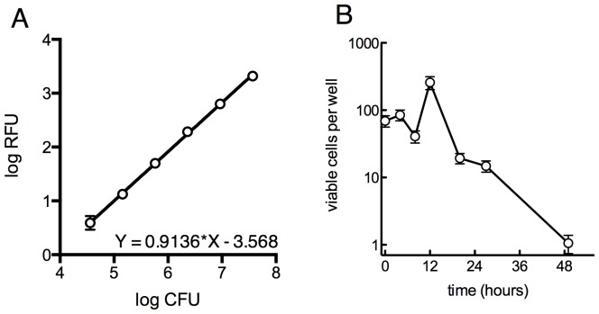
Figure 1—figure supplement 1. Survival of overall bacterial population during selection and determination of limit of detection for outgrowth of a well.

Figure 1—figure supplement 1.

(A) Determination of the limit of detection by fluorescence. GFP-expressing M. smegmatis was serially diluted in 4-fold steps and 30 µl was distributed into a 384-well plate and read as above. Samples were plated on solid medium to determine the CFU per well. Approximately 105 cells are required for a signal 10 units above background. Average of 4 replicates ± SD. (B) Viable cells per well over the first 48 hr of treatment as measured by MPN, ±95% CI.
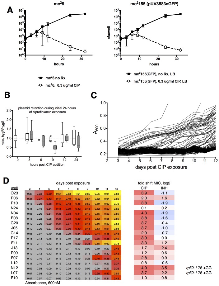
Figure 1—figure supplement 2. Ribosomal mutants can be isolated from a true wild-type M. smegmatis strain (mc26), demonstrating that the fluoroquinolone resistance of ribosomal mutants is not linked to the highly transformable mutant phenotype of mc2155.

Figure 1—figure supplement 2.

(A) Kill kinetics of M. smegmatis mc26 (left) and mc2155(pUV3583cGFP) after exposure to 0.3 µg/ml ciprofloxacin in 384-well plates in the absence of hygromycin selection. Killing was assessed at each of the timepoints shown on the x-axis by harvesting eight wells, which were then serially diluted in four fold steps, after which 5 µl of each dilution were spotted onto rectangular petri dishes; colonies were counted after 2 and 3 days of growth. Error bars indicate the standard deviation. Outliers were not excluded from this analysis. The nearly identical kill kinetics of the two strains as assayed by cfu formation on non-selective growth medium confirms that the outgrowth of GFP positive wells when mc2155(pUV3583cGFP) is exposed to ciprofloxacin in the absence of hygromycin selection is unrelated to differential plasmid retention by this highly transformable strain. (B) Assessment of plasmid loss in mc2155(pUV3583cGFP) during the time period shown in A. Colonies were plated on both LB and LB containing 50 µg/ml hygromycin B. The box-and whisker plot shows the ratio of hyg-resistant to hyg-sensitive colonies at each time point for the eight wells harvested. Open boxes are the untreated mc2155(pUV3583cGFP), and shaded boxes are mc2155(pUV3583cGFP) exposed to 0.3 µg/ml ciprofloxacin at t = 0. Whiskers indicate 5–95% CI. Plasmid stability appears to be only slightly affected in the presence of ciprofloxacin, contradicting the possibility that plasmid loss played a role in the pattern of outgrowth observed in the original selection depicted in Figure 1. (C) Outgrowth of M. smegmatis mc26 ciprofloxacin-resistant mutants under section in 0.3 µg/ml ciprofloxacin over 12 days. A total of 60 wells showed detectable fluorescence by day 12, yielding a mutation frequency of 1.86 × 10⁻⁵ based on the peak population of approximately 4.4 × 10³ bacteria per well observed 6 hr after initial exposure to ciprofloxacin in panel A. (D) Outgrowth, phenotypic, and genotypic characterization of selected wells. Outgrowth during the assay is indicated by increasing A600nm and is heatmapped to highlight the timing and intensity of growth. After 12 days, representative wells were harvested and further expanded in 4 ml cultures with 0.3 µg/ml CIP prior to assaying for ciprofloxacin and INH sensitivity. PCR sequencing of mutants N12 and L07 revealed identical GG insertions in rplO, resulting in a frameshift and premature truncation of the protein. The lfrR open reading frame and gyrA QRDR were unchanged in these mutants.