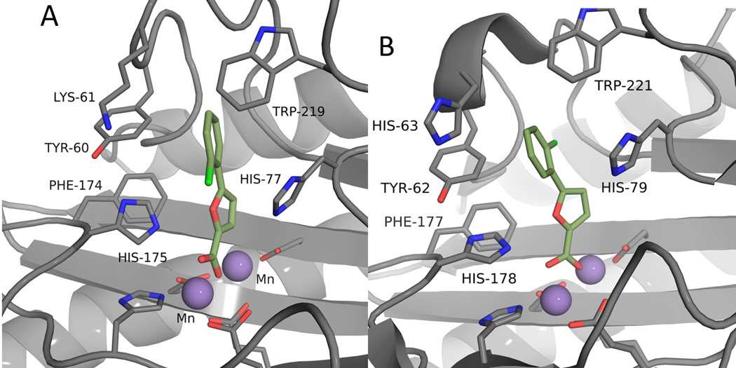
Figure 3.
Comparison of docked and observed binding interactions of (10). A: Docked pose of (10) with RpMetAP (PDB: 3MX6). B: Crystal structure of (10) bound to EcMetAP (PDB: 1XNZ).12 Note that residue numbering differs by two between the two MetAP species (Ex: RpMetAP Tyr60 and EcMetAP Tyr62 are equivalent).

