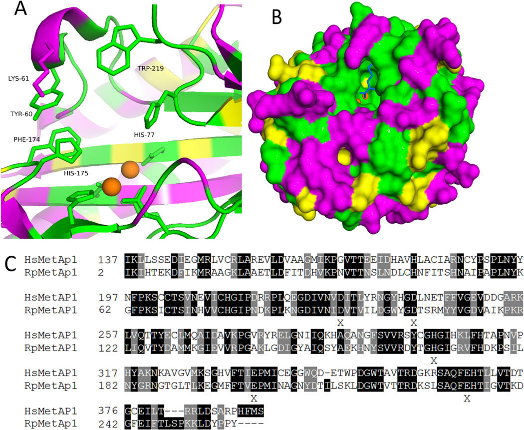
Figure 4.
Comparison of HsMetAP1 and RpMetAP1 sequences. A: Active site of RpMetAP1 colored according to sequence alignment (green = identity; yellow = similarity; magenta = non-conserved). B: Surface diagram of RpMetAp1 colored according to sequence alignment (green = identity; yellow = similarity; magenta = non-conserved). The blue ligand is a bound methionine and orange spheres are manganese cofactors. C: Sequence alignment of RpMetAP1(PDB: 3MX6) and HsMetAP1 (PDB: 2B3K) shaded according to alignment (dark = identity; light = similarity; no-shading = non-conserved). Alignment reveals 43% identity between the proteins. Residues responsible for cofactor binding are marked as X.

