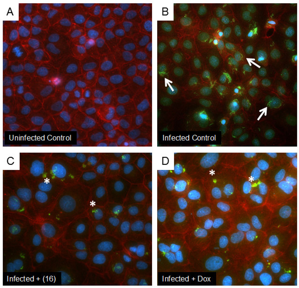
Figure 6.
Representative fluorescent micrographs showing effects of RpMetAP inhibitor compound (16) compared to a doxycycline control. Panel A shows uninfected control ECs. Panel B shows ECs infected with R. prowazekii. Panel C shows the effects of treating R. prowazekii-infected ECs with compound (16) (300 µM final concentration). Panel D shows the effects of treating R. prowazekii-infected ECs with doxycycline (25 µg/mL) as a positive control. Uninfected control cells show very little background antibody staining. The infected control shows infection with many bacilli (white arrows). Treatment with either inhibitor caused bacterial shape changes consistent with condensation and death (white asterisk). Nuclei were stained with Hoescht (blue pseudocolor); actin was stained with Texas Red-labeled Phaloidin (red pseudocolor); and rickettsiae were stained with a specific antibody conjugated to FITC (green pseudocolor).

