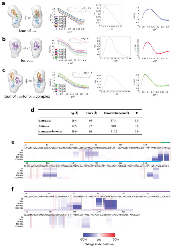
Extended Data Figure 6. Hybrid structural analysis of human Izumo1 and Juno in a solution state.
Ab initio small-angle X-ray scattering (SAXS) reconstruction, experimental scattering curves, normalized pair distance distribution function, P(r) and Kratky plot showing the degree of flexibility of (a) Izumo122-254, (b) Juno20-228, and the (c) Izumo122-254-Juno20-228 complex. No concentration-dependent or radiation effects were observed in the SAXS data. The inset box in the experimental scattering data shows linearity in the Guinier plot at low q (qRg <1.3). The Izumo122-254, Juno20-228 and Izumo122-254-Juno20-228 complex crystal structures were docked into the SAXS reconstructed molecular envelopes. The boomerang shape and upright conformation seen in the crystal structures of unbound and bound Izumo122-254, respectively, were recapitulated by the SAXS reconstructions. (d) Summary of the experimentally derived SAXS parameters for Izumo122-254, Juno20-228 and Izumo122-254-Juno20-228. The program SCATTER47 was used to calculate the radius of gyration (Rg), maximum linear dimension (Dmax), and to perform Porod-Debye analysis to obtain the Porod volume and P coefficient. Comparative deuterium exchange mass spectrometry (DXMS) profile of unbound and bound (e) Izumo122-254 and (f) Juno20-228. The plots reveal the change in individual deuterium exchange for all observable residues. The coloured lines above the residue numbers correspond to the observed regions in the crystal structures.

