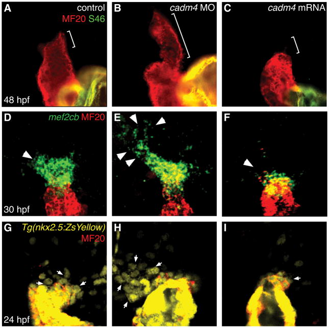
Figure 3.
The adhesion molecule Cadm4 limits OFT size by restricting SHF progenitor production. (A–C) Hearts at 48 hpf, stained with MF20 (red) and S46 (green) antibodies (Alexander et al., 1998) to visualize the OFT (red, bracket), ventricle (red), and atrium (yellow). Compared to controls (A) with normal OFT length, cadm4 morphants (B) exhibit a significantly elongated OFT, and embryos injected with cadm4 mRNA (C) have a significantly reduced OFT. (D–F) In situ hybridization shows expression of mef2cb (green) in SHF progenitor cells adjacent to the differentiated myocardium at the arterial pole of the heart tube (MF20, red); dorsal views at 30 hpf. In comparison with controls (D), the SHF progenitor population (arrowheads) is expanded in cadm4 morphants (E) and reduced in embryos overexpressing cadm4 (F). (G–I) Dorsal views show Tg(nkx2.5:ZsYellow) (Zhou et al., 2011) expression at 24 hpf; MF20 (red) marks differentiated cardiomyocytes. Controls (G) exhibit a normal number of SHF progenitors (arrows) in the region proximal to the arterial pole. cadm4 morphants (H) exhibit a significant surplus of progenitors in this region, and embryos overexpressing cadm4 (I) exhibit a significant reduction of progenitors. Adapted from Zeng and Yelon (2014).

