Figure 8.
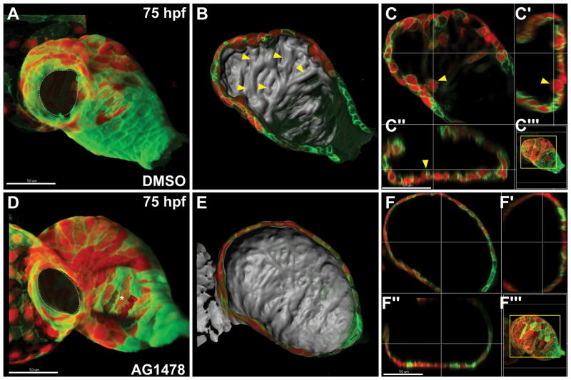
Neuregulin signaling is required for the initiation of trabeculation. Confocal reconstructions of the ventricular myocardium in wild-type embryos, expressing Tg(myl7:dsredt4) and Tg(myl7:egfp-hshras) and treated either with DMSO (A–C) or AG1478 (D–F), an inhibitor of ErbB receptors, from 27–75 hpf. Volume reconstructions (A,D) and lumenal surface reconstructions (B,E) are shown. The AVC is outlined with a dotted white line (A,D). White asterisk (D) indicates an area where the acquisition of fluorescent signal was blocked by overlying melanocytes. Optical slices show sagittal sections (C, F), transverse sections (C′, F′), and coronal sections (C″, F″) through the region of interest, as shown in maximum intensity projection (C‴, F‴). (A–C) DMSO-treated control embryos exhibit lumenal protrusions and primitive ridges (yellow arrowheads). (D–F) In contrast, AG1478-treated embryos do not initiate trabeculation and instead exhibit a smooth lumenal surface and a uniform thickness of the chamber wall. Adapted from Peshkovsky et al. (2011).

