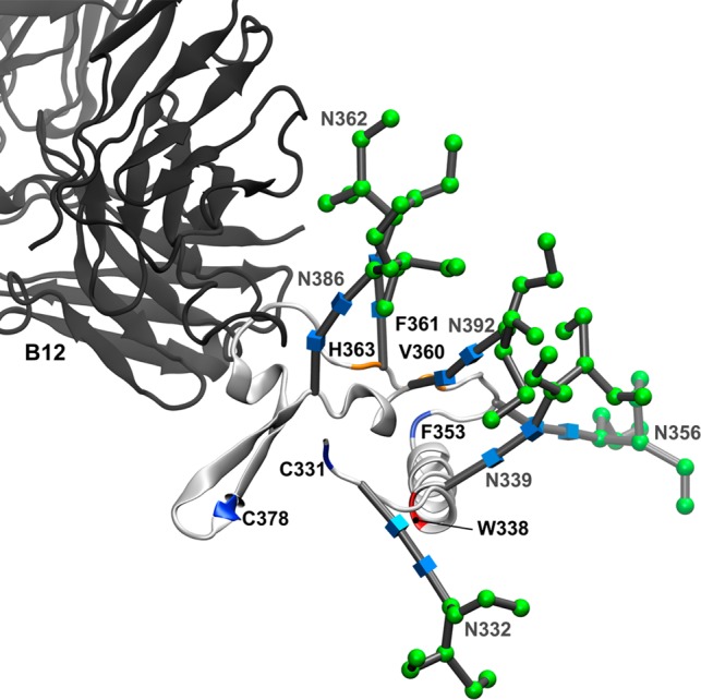
Figure 8.

Interaction of b12 with the C3 domain of gp120 mediated by N-linked glycosylation. Backbone and N-linked glycans of the C3 domain from the full-length, glycosylated gp120 MD simulation (light gray ribbon) aligned with the b12 Fab fragment (dark gray ribbon). All other gp120 domains have been excluded for the sake of clarity. Residues that showed >80% protection from HR-HRPF upon b12 binding are colored red. Residues that showed between 40 and 80% protection from HR-HRPF upon b12 binding are colored orange. Residues showing no protection from HR-HRPF upon b12 binding are colored blue. Glycans are labeled and shown as 3D-SNFG symbols, which are positioned at each residue’s ring center.
