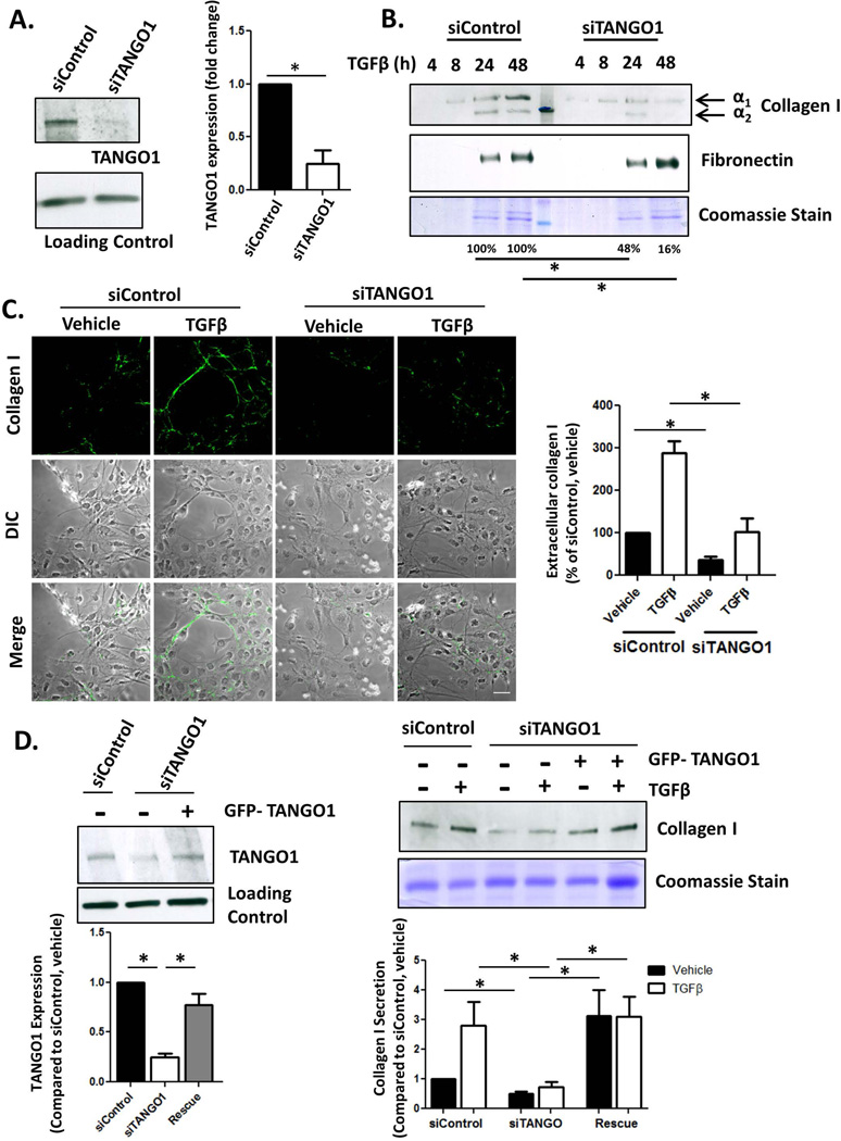
Figure 1. TANGO1 is critical for collagen I secretion from HSCs.
A. LX-2 cells were transfected with an siRNA against TANGO1 (siTANGO1) or a scrambled Control (siControl). 72hrs after transfection, TANGO1 expression was analyzed by Western Blot with HSC70 serving as a loading control (quantification in the adjacent graph, *=p<0.05). B. siControl or siTANGO1 LX-2 cells were treated with 10ng/mL TGFβ or a vehicle control for 24hrs. Conditioned media was harvested, separated via SDS-PAGE, and collagen I and fibronectin levels were analyzed using immunoblotting (upper panel, *=p<0.05). Coomassie stain was used to confirm equal loading between siControl and siTANGO1 pairs since conditioned media was harvested at different time points (lower panel). Quantification is displayed below the blots, n=4, *=p<0.05. C. Confocal images were taken of extracellular collagen I deposition by siControl or siTANGO1 LX-2 cells treated with vehicle or TGFβ. Cells were fixed but not permeabilized in order to eliminate staining for intracellular procollagen I. Pixel intensity was determined using ImageJ, adjusted for cell number, and is displayed in the adjacent graph. (Scale bar=50µm, n=4, *=p<0.001). D. LX-2 cells were cotransfected with an siRNA against TANGO1 (or control siRNA) and a plasmid expressing exogenous TANGO1 (or a control plasmid) for 48 hours followed by TGFβ treatment for 24hr. Whole cell lysate (Left Panel) was harvested and immunoblotted for TANGO1 and HSC70 (Loading Control). Conditioned media was also harvested, separated by SDS-PAGE and collagen I secretion was examined by immunoblotting (Right Panel). Coomassie staining confirmed equal loading. N=5, *=p<0.05.

