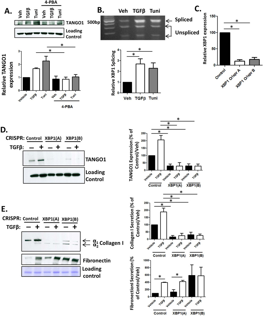
Figure 5. The UPR drives expression of TANGO1.
A. siTANGO1 or siControl transfected LX-2 cells were serum starved in basal media containing 4-PBA or a vehicle for 4hr, followed by treatment with TGFβ or tunicamycin (Tuni) for 24hr. After treatment, cells were harvested, lysed, and analyzed for TANGO1 expression by immunoblotting. Quantification is displayed below (n=4, *=p<0.01). B. hHSCs were treated with vehicle or TGFβ and analyzed for XBP1 cleavage. Unspliced XBP1 is digested into two fragments (290bp and 183bp) and spliced XBP1 is visualized at 473b. Quantification located below, n=5, *=p<0.001. C. LX-2 cells were stably infected with virus expressing one of two CRISPR constructs (A or B) targeting XBP1 or a control construct. XBP1 expression was analyzed by qPCR. D and E. Cells lacking XBP1 or control cells were treated with TGFβ or vehicle, and TCL and conditioned media were harvested. TANGO1 expression (TCL, D, n=3, *=p<0.01) and collagen I or fibronectin secretion (conditioned media, E, n=4, *=p,0.05) were measured using immunoblotting, following by ImageJ analysis in the adjacent graphs.

