
Fig. 1.
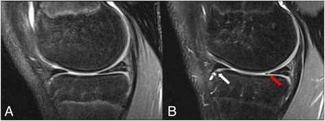
Sagittal PD FS images showing the medial compartment of the femorotibial joint of an adolescent male volleyball player at baseline a and 2- year follow-up b. In the follow-up scan, there is a new osteophyte at the anterior cortex of the tibial plateau (white arrow) and a new vertical tear of the posterior horn of the meniscus (red arrow), which were not visible in the baseline scan