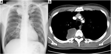
Fig. 1.

Radiologic imaging of the chest of a 48-year-old man with pleomorphic carcinoma of the lung. a Chest radiograph reveals a 6-cm shadow in the right upper lung field. b Chest computed tomography reveals a 6.5-cm tumor in the right upper lobe with invasion to the chest wall
