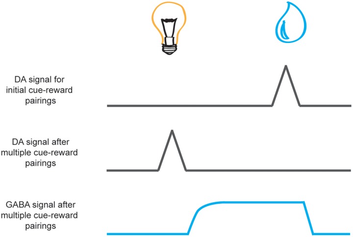
FIGURE 3.
Schematic of dopamine and GABA reward prediction-error activity during learning. Neural activity is aligned to cue presentation (e.g., light, on the left) and reward presentation (e.g., a drop of juice, on the right). While phasic activity of dopamine neurons (black lines) are elicited by unexpected reward delivery upon initial cue-reward pairings (top) with repeated cue-reward pairings the signal at the time of reward receipt wanes as the reward becomes predicted by the cue (middle). This transition occurs gradually over successive trials in accordance with traditional learning models of prediction error (Rescorla and Wagner, 1972; Sutton and Barto, 1981). It is speculated that this reduction in the dopamine signal to the reward may result from inhibition of dopamine neurons by GABAergic neurons in the VTA (bottom, blue line) that is initiated after cue offset and persists during reward delivery (Houk et al., 1995; Cohen et al., 2012).

