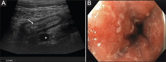
Figure 2.

(A) Cross-sectional image of acute Crohn’s colitis in the sigmoid colon (9 MHz probe) Blurred stratification of the thickened bowel wall (arrow) and marked fibro-fatty proliferation (asterisk). (B) Corresponding endoscopic view of the same patient during the same week
