
Figure 1.
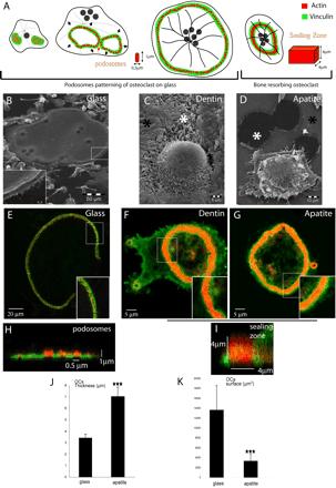
Formation of podosome belt in spread osteoclasts or sealing zone in apico-basal polarized osteoclasts is dependent on extracellular matrix. (A) Scheme of the different actin structures observed in osteoclasts. Osteoclasts seeded on glass form podosomes, small cylinders of actin surrounded by vinculin. Podosomes organize into three different structures along differentiation namely clusters, rings, and belts into mature osteoclasts (Destaing et al., 2003). On bone when resorbing, they form a sealing zone, a large circular band of actin surrounded by vinculin. Differentiated osteoclasts were plated on either glass (B, E, and H), dentin (C–F), or apatite (D, G, and I); scanning electron microscopy images of osteoclasts adherent on their respective substratum are shown (B–D). Mature osteoclasts adherent on glass are large flat cells with a swollen area at site of podosome belt (B, inset). In contrast, on dentin or apatite substrate-resorbing osteoclasts are contracted. Black asterisks indicate nonresorbed matrix and white asterisks resorption pits (C and D). (E) A single osteoclast on a glass coverslip is shown with a podosome belt; the inset is a zoom of the area outlined to show podosomes (bar, 20 μm). (F and G) Osteoclast on dentin and apatite exhibiting a sealing zone; inset is a zoom of the area outlined showing the absence of podosomes and the presence of a large sealing zone (bar, 5 μm). (H and I) Z-cuts of podosome belt and sealing zone. Actin is shown in red and vinculin in green. (J and K) Graphical representation of surface area and thickness of osteoclasts adherent on either glass coverslips or apatite-coated glass coverslips, measured using Zeiss LSM 510 software. Stars indicate statistically significant results using a Student' t test (p < 0.0001; n = 30).