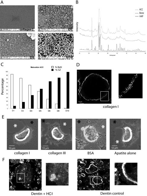
Figure 3.
Apatite mineral triggers sealing zone formation. (A) Scanning electron microscopy showing the nature of the coating formed along the mineralization process of ACC slides. Apatite particles appeared on day 1, grew, and covered the entire surface by day 5. On day 10, three-dimensional bone-like apatite structures were formed. (B) X-ray diffraction patterns for the substrate ACC in the range of 20–90° 2 theta is presented. As can be seen, ACC substrate showed the characteristic peaks of apatite (002, 310) with some unresolved large broad peaks typical of a poorly crystalline apatitic calcium phosphate. By comparison of the ACC diagram with those of bone samples and hydroxyapatite, we could see that, if the same apatitic phase is common to all the samples, bone as ACC showed a poorly crystalline diagram, compared with hydroxyapatite, which logically presents a highly crystalline phase. (C) Mature osteoclasts were seeded on coverslips at different time points during the mineralization process and stained for actin 24 h later. The percentage of osteoclasts exhibiting podosome belts vs. sealing zones along the mineralization process is shown (n = 200 at each day). (D and E) Whereas osteoclasts seeded on collagen I exhibited podosome belts, they formed sealing zone as long as organic coating (collagen I, collagen III or BSA) was mineralized. (F) In contrast to dentin, osteoclasts adherent on HCl demineralized dentin slices did not form clearly recognizable actin structures.

