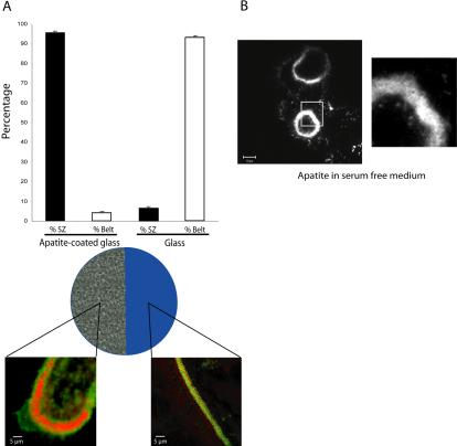
Figure 4.
Sealing zone formation is not triggered by microenvironmental modifications. (A) To further confirm the role of extracellular matrix on osteoclast actin organization, we used apatite-coated slides. Half the slide was first immersed in HCl (6 N) to remove apatite crystals. Mature osteoclasts were then seeded on these slides for 24 h. Osteoclasts exhibit sealing zones when adherent on the mineral part and podosome belts on glass. The graph represents the percentage of osteoclasts exhibiting podosome belts or sealing zones according to the substratum (n = 150). (B) Mature adherent osteoclasts still exhibit a sealing zone on apatite in serum-free medium ruling out the putative role of medium components.

