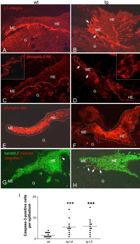
Figure 7.
Altered β1-integrin expression and FAK phosphorylation, reduced Akt phosphorylation, and activation of apoptotic signaling in 5-d wounds of transgenic mice. Immunofluorescence staining of frozen (C, D, G, and H) or ethanol/acetic acid-fixed paraffin sections (A, B, E, and F) from 5-d old wounds of wild-type and transgenic animals. G, granulation tissue; HE, hyperproliferative epithelium; ME, migrating epithelium. (A and B) Staining for β1-integrins; scattered keratinocytes are marked with arrowheads. (C and D) staining for phospho-FAK (Y397). A higher magnification of the epithelial tongue (1000×) is shown in the corners of the pictures. (E and F) Staining for phospho-Akt; the borders of the epithelium are marked with a white line. (G and H) Staining for cleaved caspase-3 (red) and type II-keratin (green); apoptotic keratinocytes (yellow) are marked with arrowheads. (I) Number of caspase-3–positive cells per epithelium in wild-type and transgenic animals of two different founder lines. Wound sections were counted regardless of the severity of their phenotype. Statistical analysis was performed using GraphPad Prism4 software. The horizontal line indicates the median (the 50th percentile), and each dot corresponds to one analyzed epithelium. p values are p = 0.0006 for comparison of wt with line 4 and p = 0.0002 for comparison of wt with line 3, according to Student's t test.

