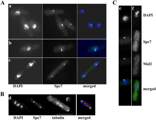
Figure 2.
Spc7 localizes to the kinetochore. (A) Localization of the Spc7-GFP protein in wild-type cells in interphase (a) early (b) or late (c) stages of mitosis. Fixed cells were stained with DAPI and anti-GFP antibody. (B) Localization of the Spc7 fusion in a wild-type cell arrested by overexpression of mph1+. GFP signals; spindles and condensed chromosomes were simultaneously observed by staining with anti-GFP antibody, anti-tubulin antibody and DAPI. (C) Colocalization of Spc7-HA (green) and Mal2-GFP (red) fusion proteins in interphase cells (e) and cells arrested by overexpression of mph1+ (f). Chromosomes, HA- and GFP-signals were simultaneously observed by staining with anti-GFP antibody, anti-HA antibody, and DAPI. Bar, 10 μm.

