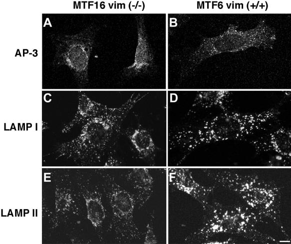
Figure 6.
AP-3 and lysosomal antigen distribution are modified in fibroblasts from vimentin-deficient mice. MTF16 vimentin –/– and MTF6 vimentin +/+ cells were fixed and processed for immunofluorescence confocal microscopy. Cells were stained with antibodies to the AP-3 δ subunit (a and b), LAMP I (c and d), or LAMP II (e and f). As in SW13 cells, in fibroblasts lacking vimentin, AP-3 and lysosomal antigens are repositioned to the juxtanuclear region. Additionally, we observed differences in the size and intensity of LAMP-positive organelles. All experiments were done in duplicate coverslips on at least two independent experiments, and five to 10 random fields were imaged per coverslip.

