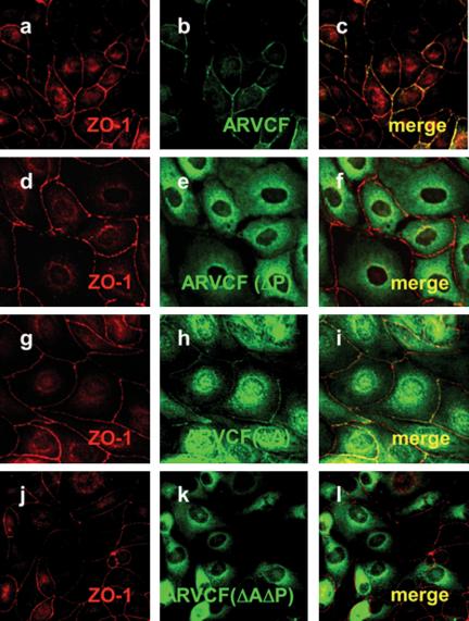
Figure 3.
Colocalization of ARVCF and ZO-1 in transfected MDCK cells. MDCK cells transfected with wild-type ARVCF (a-c) or mutant ARVCF(ΔP) (d-f), ARVCF(ΔA) (g-i), or ARVCF(ΔAΔP) (j-l) were grown on glass coverslips, fixed, permeabilized, and stained for ZO-1 (red, a, d, g, and j) and ARVCF (green, b, e, h, and k). Confocal images of the ZO-1 and ARVCF labeling were merged to show regions of colocalization (yellow, c, f, i, and l).

