Figure 3.
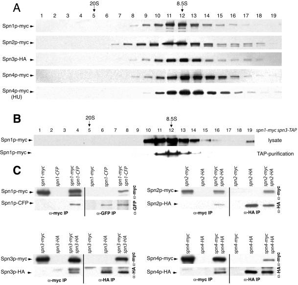
Characterization of S. pombe septin complexes. (A) Lysates prepared in NP-40 buffer from each myc-epitope– or HA-epitope–tagged septin protein from asynchronously growing cells or a hydroxyurea (HU)-arrested culture were resolved on sucrose gradients. Fractions were collected from the bottom of the gradient (1) and immunoblotted with 9E10 or 12CA5 to detect myc or HA-tagged proteins. The peaks of thyroglobulin (20S) and aldolase (8.5S) collected from gradients prepared and run in parallel are indicated. (B) A TAP and a lysate prepared in NP-40 buffer from spn1-myc spn3-TAP cells were resolved on sucrose gradients. Fractions were collected from the bottom of the gradient and immunoblotted with 9E10 to detect Spn1p-myc. The peaks of thyroglobulin (20S) and aldolase (8.5S) collected from gradients prepared and run in parallel are indicated. (C) anti-myc (left side of panels) and anti-GFP or anti-HA (right side of panels) immunoprecipitates from the indicated strains were blotted with anti-myc (top panels) and anti-GFP or anti-HA (bottom panels) antibodies.

