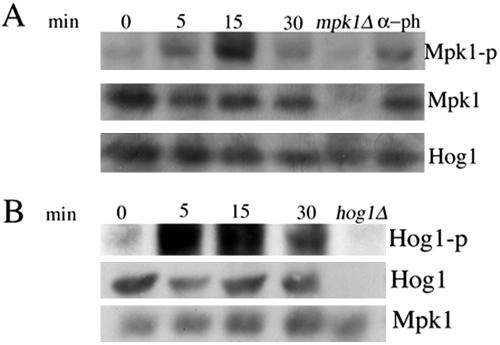
Figure 10.
(A) Mpk1 is phosphorylated on exposure of cells to hydrogen peroxide. Mid-log cultures of wild-type strain cells (EY957) were treated with hydrogen peroxide at 5 mM final concentration with 50 nM α-pheromone as a positive control. Samples were taken at the indicated times (0, 15, and 30 min). Wild-type cells were treated with α-pheromone for 60 min. Hydrogen peroxide was added to mpk1Δ strain for 15 min. Western blot analysis was performed as described in Materials and Methods by using anti-phospho-p44/p42 antibody to detect the phosphorylated Mpk1, Kss1 and Fus3, anti-Mpk1 to detect the total protein and anti-Hog1 as a loading control. (B) Hydrogen peroxide activates Hog1 MAPK. Mid-log cultures of wild-type strain cells (EY957) were treated with hydrogen peroxide at 5 mM final concentration. Samples were taken at the indicated times (0, 15, and 30 min). Strain disrupted in HOG1 gene, used as negative control, was treated with hydrogen peroxide for 15 min. Western blot analysis was performed as described in Materials and Methods by using anti-phospho-p38 antibody to detect the phosphorylated Hog1, with anti-Hog1 to detect the total protein and anti-Mpk1 as a loading control.

