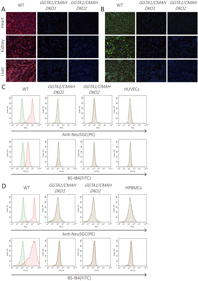
Fig. 4.
Immunofluorescence and flow cytometry analysis of Gal and Neu5Gc expression in GGTA1/CMAH DKO pigs. (A–B) Immunofluorescence of various tissues from wild-type or GGTA1/CMAH DKO pigs stained with anti-Neu5Gc antibody (A) and BS-IB4 lectin (B) (Magnification × 200; Neu5Gc, red; Gal, green; Nuclei, blue). (C–D) Flow cytometry analysis of AECs (C) and PBMCs (D) from the pigs indicated in A-B stained with anti-Neu5Gc antibody and BS-IB4 lectin. Human umbilical vein endothelial cells (HUVECs) and human PBMCs (HPBMCs) were negative controls, respectively. The green lines represent negative controls; the red lines represent the experimental groups.

