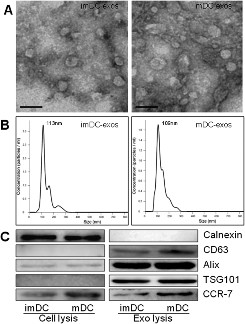
Figure 1. Successful isolation of exosomes from DCs culture medium.
(A) The ultrastructure of exosome by transmission electron microscopy. Bar size, 100 nm. (B) The size distribution profile of immature and mature DC-exos by Nanosight, revealing a size peak of 113 nm in immature DC-exos and 109 nm in mature DC-exos. (C) The expression of exosomes negative marker, Calnexin and positive markers, Alix, CD63 and TSG101. And also, the expression of CCR7 was detected in both cell lysis and exosomes lysis. A total of 20ug protein from DCs lysis and 5 ug protein from exosomes lysis was loaded into each lane. Full-length blots can be found in Supplemental Figures 3 and 4.

