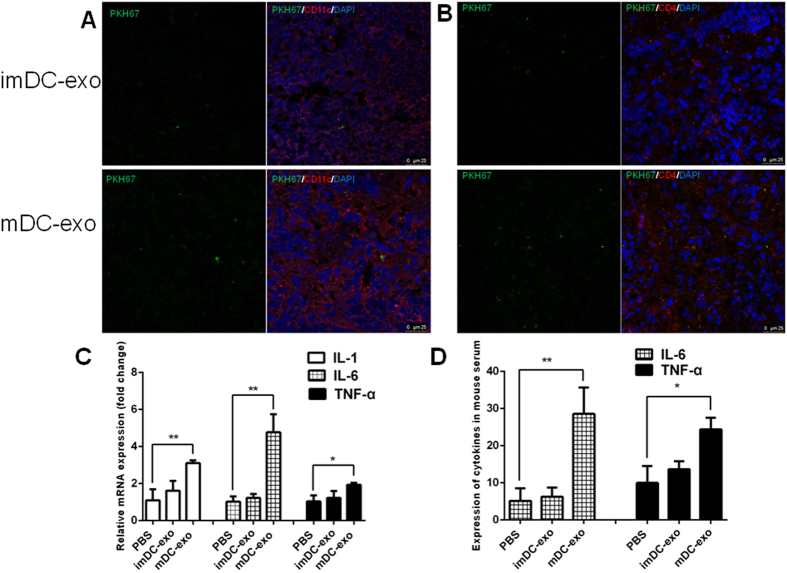
Figure 5. DC-exos uptake by splenetic DCs and T cells and inducement of inflammation in vivo.
(A,B) The PKH67-labelled immature and mature DC-exos were intravenously injected into mice. After 24 hours, the mouse was sacrificed and the spleen was cut into 6um sections for staining CD11c (A) and CD4 (B) and DAPI. The uptake of DC-exos by splenetic DCs and T cells was observed under a confocal microscope. Representative images of 3 dependent studies. (C) Twenty-four hours after PBS or DC-exos injection, the spleen was harvested and the tissue total RNA was extracted for detection of IL-1, IL-6 and TNF-α by quantitive PCR (n = 4–5). (D) Twenty-four hours after PBS or DC-exos injection, the serum was collected for detection of IL-6 and TNF-α by Elisa (n = 5–6). *p < 0.05, **p < 0.01, ***p < 0.001.

