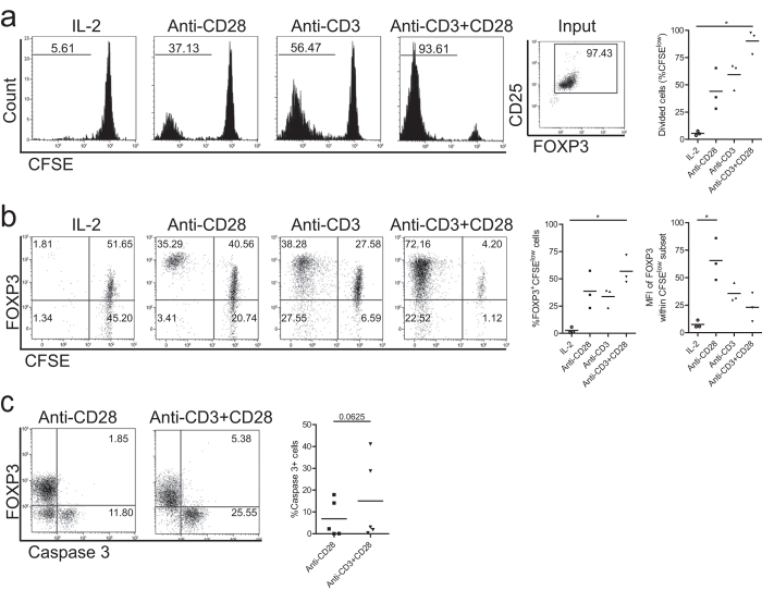
Figure 1. Single-CD28 stimulation induces Treg proliferation and promotes high level FOXP3 expression.
Flow cytometry of FACS-sorted human Treg (CD4+CD25high) that were labeled with CFSE and stimulated with soluble CD28 mAb, plate bound CD3 mAb or both (Anti-CD3+CD28) in the presence of rhIL-2. As a control Treg cultured in the presence of rhIL-2 only were included. Cell division indicated by the dilution of CFSE (a), intracellular expression of FOXP3 (b) and active caspase 3 (c) were determined at day 7 of the cultures. Numbers within the histograms indicate the percentage of divided cells (a) and numbers within the quadrant show the percentage of positive cells (b,c). Cumulative data of Treg proliferation (a, right panel), the median fluorescence intensity (MFI) of FOXP3 (b, right panel), and the percentage of apoptotic cells (c) right panel) are also shown. Bar in cumulative data indicates the mean value. Kruskal-Wallis followed by Dunns post-hoc test (a) n = 3; b, n = 3) and Wilcoxon signed-rank test (c, n = 5) were used for statistical analysis. *P < 0.05.

