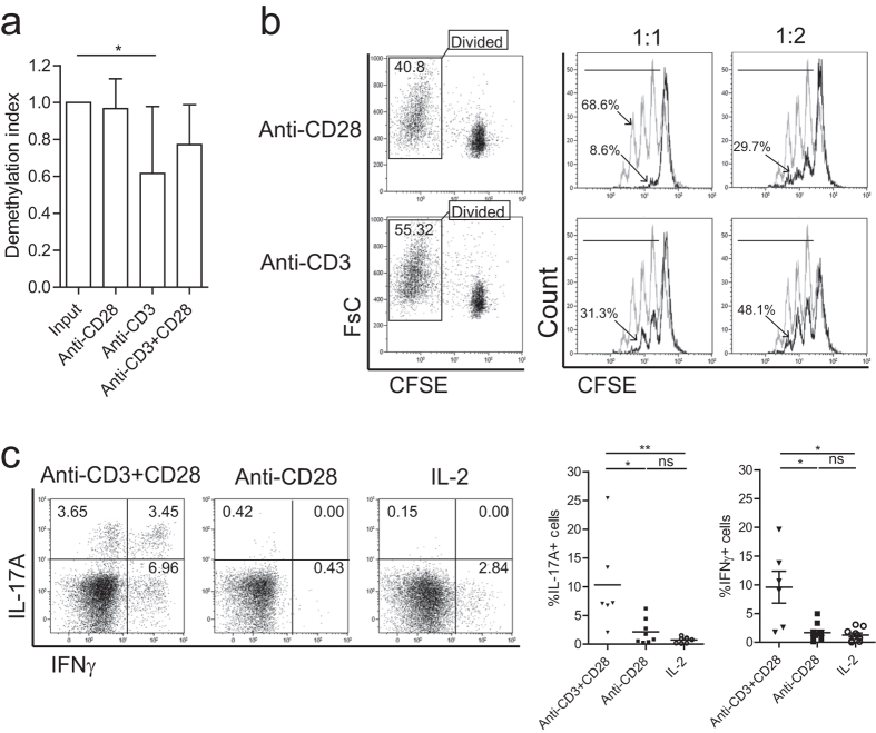
Figure 2. Single-CD28 stimulated Treg reveal a highly demethylated FOXP3 gene and profound suppressor function.
(a) FACS-sorted human Treg were stimulated with soluble CD28 mAb, plate bound CD3 mAb or both (anti-CD3+CD28) in the presence of rhIL-2 for 7-days. Thereafter, cells were harvested and the demethylation status of FOXP3 gene was analyzed using bisulfate sequencing. n = 6–7. (b) CFSE-labeled FACS-sorted Treg were stimulated as described above. The divided cells (CFSE low population) were re-sorted (left panel) at day 7 of the cultures, and subsequently their suppressive function was analyzed in a co-culture suppression assay. Overlay histograms show the inhibition of responder T cells (Tresp) proliferation following the addition of graded doses of Treg. Numbers indicate the percentage of divided responder T cells. Grey line: stimulated Tresp, Black line: co-cultured with Treg of interest. The ratio of Treg:Tresp are indicated on the top. Representative experiment of n = 3 individual experiments conducted with cells obtained from different donors are shown. (c) Intracellular staining of cytokine IL-17A and IFNγ after additional stimulation with PMA, ionomycin, and Brefeldin-A for 4 hours. Numbers within the quadrant indicate the percentage of positive cells. Dotplots show a representative experiment of n = 6–10 individuals as shown in the cumulative data graph (right panel). Kruskal-Wallis followed by Dunns post-hoc test was used for statistical analysis (a,c). *P < 0.05; **P < 0.01; ns: not significant.

