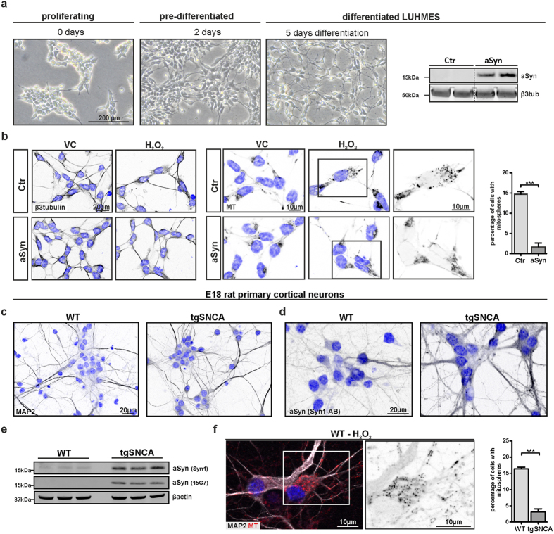
Figure 5. Increased aSyn expression reduces the formation of oxidative stress-induced mitospheres in neurons.
(a) Proliferating, pre-differentiated and differentiated LUHMES cells visualized via light microscopy are depicted. aSyn (15G7) levels were detected by WB in differentiated Ctr and aSyn LUHMES cells. Bands shown for Ctr and aSyn LUHMES samples are derived from the same WB. (b) Differentiated LUHMES cells were treated with 50 μM H2O2 for 4 h and stained for β3-tubulin and with MT. Quantification of the percentage of cells with mitospheres revealed that aSyn overexpression prevented mitosphere formation in LUHMES cells (unpaired two-tailed t-test, n = 3 with >160 cells quantified per sample, p = 0.0004). (c–f) E18 rat primary cortical neurons of wild type (WT) or transgenic animals overexpressing human aSyn (tgSNCA) were cultured for 8 days in vitro (DIV8). (c) Immunocytochemical stainings for the neuronal marker MAP2 and (d) for aSyn (Syn1 antibody, detects human and rodent aSyn) are depicted. (e) WBs of aSyn detected via an antibody for both rodent and human aSyn (Syn1), as well as a human specific antibody (15G7) is shown. (f) Overexpression of human aSyn reduced mitosphere formation in cultures of primary cortical neurons, as quantified for cells treated with 200 μM H2O2 for 4 h (unpaired two-tailed t-test, n = 3 with >150 cells quantified per sample, p = 0.0002).

