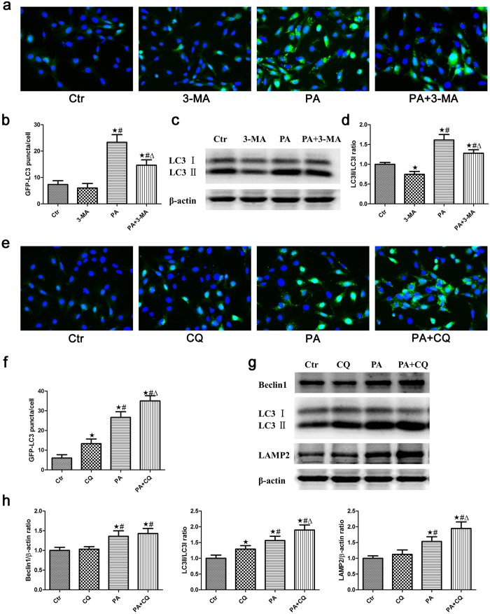
Figure 2. The effect of palmitic acid on autophagic flux in podocytes.
(a) Podocytes stably expressing GFP-LC3 were pretreated with 2 mmol/L 3-MA for 1 hour, treated with 150 μmol/L palmitic acid for 24 hours and then analyzed by fluorescence microscopy (400×). (b) GFP-LC3 puncta (mean ± SEM) were quantified for each experiment (n = 3). At least 30 cells were counted in each individual experiment. ∗p < 0.05 vs. control group, #p < 0.05 vs. 3-MA group, Δp < 0.05 vs. PA group. (c) Podocytes were treated with 150 μmol/L palmitic acid in the absence or presence of 2 mmol/L 3-MA for 24 hours, and the cell lysates were then analyzed by immunoblot using an antibody against LC3. (d) Densitometric analysis of LC3-II/LC3-I expression in Figure c. ∗p < 0.05 vs. control group, #p < 0.05 vs. 3-MA group, Δp < 0.05 vs. PA group. (e) Podocytes stably expressing GFP-LC3 were pretreated with 3 μmol/L CQ for 1 hour, treated with 150 μmol/L palmitic acid for 24 hours and then analyzed by fluorescence microscopy (400×). (f) GFP-LC3 puncta fluorescence (mean ± SEM) were quantified for each experiment (n = 3). At least 30 cells were counted in each individual experiment. ∗p < 0.05 vs. control group, #p < 0.05 vs. CQ group, Δp < 0.05 vs. PA group. (g) Podocytes were treated with 150 μmol/L palmitic acid in the absence or presence of 3 μmol/L CQ for 24 hours, and the cell lysates were then analyzed by immunoblot using antibodies against Beclin1, LC3 and LAMP-2. (h) Densitometric analysis of Beclin1, LC3-II/LC3-I and LAMP-2 expression in Figure g. ∗p < 0.05 vs. control group, #p < 0.05 vs. CQ group, Δp < 0.05 vs. PA group. Ctr: Control group, podocytes were treated with 1% BSA; 3-MA: Podocytes were treated with 2 mmol/L 3-MA for 24 hours; PA: Palmitic acid group, podocytes were treated with 150 μmol/L palmitic acid for 24 hours; PA + 3-MA: Podocytes were treated with 150 μmol/L palmitic acid for 24 hours after pretreatment with 2 mmol/L 3-MA for 1 hour; CQ: Podocytes were treated with 3 μmol/L CQ for 24 hours; PA + CQ: Podocytes were treated with 150 μmol/L palmitic acid for 24 hours after pretreatment with 3 μmol/L CQ for 1 hour.

