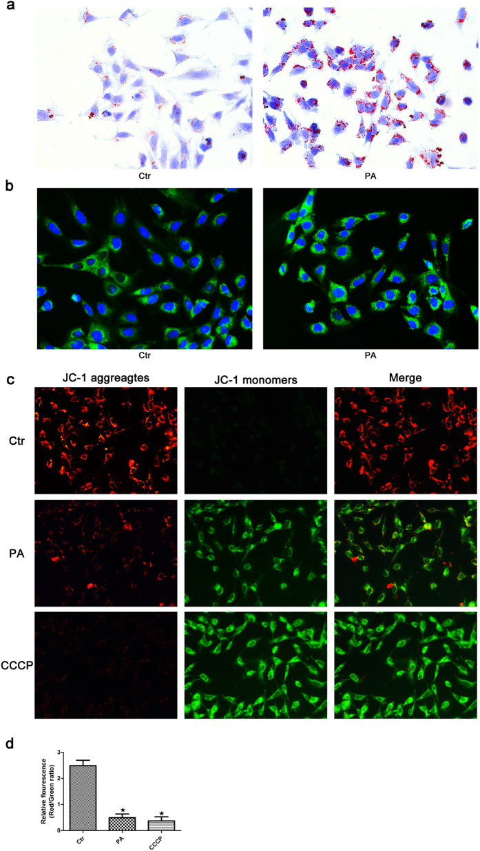
Figure 4. Palmitic acid-induced lipotoxicity promotes mitochondrial injury in podocytes.
Podocytes were treated with 150 μmol/L palmitic acid for 24 hours, and the lipid content in the cells was then detected by Oil Red O staining (400×) (a) and BODIPY lipid probes (400×) (b). (c) Palmitic acid induced a change in mitochondrial transmembrane potential (ΔΨm). Podocytes were treated with 150 μmol/L palmitic acid for 24 hours or 10 μM CCCP for 1 hour, and the cells were then stained with the fluorescent probe JC-1. CCCP, a powerful mitochondrial uncoupling agent, was used as a positive control. Red fluorescence indicates normal ΔΨm with JC-1 aggregates in mitochondria, and green reflects cytosolic JC-1 monomer indicative of ΔΨm loss (400×). (d) Fluorescent intensity from 5 randomly selected microscopic fields per group was captured and analyzed. The data are expressed as the mean ± SEM; n = 3. ∗p < 0.05 vs. control group. Ctr: Control group, podocytes were treated with 1% BSA. PA: Palmitic acid group, podocytes were treated with 150 μmol/L palmitic acid for 24 hours. CCCP: Podocytes were treated with 10 μM CCCP for 1 hour.

